



Categories
Normal Ovary Size in Women: Measurements, Changes and What They Mean
Aug 25, 2026
Key Takeaway
If you are comparing the measurements on an ultrasound report, looking at the complete findings can provide more useful context than focusing on one number alone.- A reproductive-age ovary is commonly described as around 3–5 cm in length, although individual measurements vary.
- Ovarian volume varies considerably, with research reporting values around 7 mL on average or as a median in reproductive-age women.
- The right and left ovaries do not necessarily have identical measurements.
- Small, simple ovarian cysts before menopause often do not require treatment.
- Persistent symptoms, complex cysts or unexpected ovarian enlargement should be medically assessed.
Table of Contents
- What Is Considered a Normal Ovary Size?
- Normal Ovary Size in mm and cm
- Normal Ovarian Volume (mL/cc)
- How Does Ovary Size Change With Age?
- Can Ovary Size Change During the Menstrual Cycle?
- Does Ovary Size Affect Fertility?
- What Causes Ovary Size to Change?
- Ovarian Cyst Size: Normal vs. Concerning
- When Should You See a Gynaecologist?
- Conclusion
- FAQs
What Is Considered a Normal Ovary Size?
In adults of reproductive age, ovaries are commonly described as approximately 3–5 cm long, 1.5–3 cm wide and 0.6–1.5 cm thick. These figures are broad anatomical references rather than strict diagnostic boundaries because ovarian dimensions can vary considerably between individuals. This distinction is useful when you are reading your own ultrasound report. If the right ovary is slightly larger than the left, it does not automatically mean something is wrong. Ovarian appearance can change during the menstrual cycle, and doctors consider the dimensions alongside ovarian volume, menstrual-cycle stage, follicles, symptoms and the overall ultrasound findings. So, rather than asking only, “Is one ovary larger than the other?”, it can be more useful to understand whether the difference fits with your cycle stage and the rest of the scan.1. Normal Ovary Size in mm and cm
Ultrasound reports may provide ovarian dimensions in either millimetres or centimetres. If you are comparing the measurements mentioned in your report, the following broad reference ranges can make the figures easier to understand.| Ovary | Approximate size in mm | Approximate size in cm |
| Right ovary | 30–50 × 15–30 × 6–15 mm | 3–5 × 1.5–3 × 0.6–1.5 cm |
| Left ovary | 30–50 × 15–30 × 6–15 mm | 3–5 × 1.5–3 × 0.6–1.5 cm |
2. Normal Ovarian Volume (mL/cc)
Ovarian volume combines the length, width and thickness into a single measurement. It can provide additional context when interpreting ovary size rather than relying only on ovarian length. There is no single ovarian volume that is ideal for every woman. An International Society of Ultrasound in Obstetrics and Gynaecology (ISUOG) training resource describes ovarian volume in women of fertile age as highly variable, reporting a median of about 7 mL. Recent research involving Indian women aged 18–40 found mean ovarian volumes of 7.06 cc on the left and 7.26 cc on the right with transabdominal ultrasound. With transvaginal ultrasound, the corresponding means were 7.12 cc and 7.45 cc. Therefore, a value that is slightly higher or lower than an average should not be interpreted as a diagnosis on its own. Your age, ultrasound method and overall ovarian appearance also matter.How Does Ovary Size Change With Age?
Ovarian volume generally increases through childhood and adolescence, peaks in early adulthood and gradually declines with age, particularly towards menopause. This is why age is important when interpreting ultrasound measurements.Can Ovary Size Change During the Menstrual Cycle?
Yes. Follicle growth, ovulation and corpus luteum formation can temporarily change the size and appearance of an ovary, so measurements may differ depending on when the scan is performed.Does Ovary Size Affect Fertility?
Ovary size alone cannot determine fertility or egg reserve. Age, AMH levels, antral follicle count and reproductive history usually provide more useful context when fertility is being assessed.What Causes Ovary Size to Change?
If you compare two ultrasound reports and notice that an ovary has changed in size, the difference does not necessarily mean a health problem is progressing. Ovaries are active organs, and their appearance can change over time. Factors that may influence ovarian measurements include:- Growing follicles during the menstrual cycle
- Ovulation and corpus luteum formation
- Pregnancy-related changes
- Age and menopause
- PCOS and other hormonal conditions
- Ovarian cysts
- Previous procedures involving ovarian tissue
Ovarian Cyst Size: Normal vs. Concerning
If your ultrasound also mentions a cyst, seeing the measurement alongside the ovary size can create additional worry. However, the word “cyst” does not by itself mean that surgery or serious treatment is required. A follicle normally develops within an ovary and is typically around 2–3 cm when measured across. RCOG describes a fluid-filled structure greater than 3 cm in diameter as an ovarian cyst. Many ovarian cysts that occur before menopause are simple and resolve naturally. For simple cysts in premenopausal women, RCOG provides the following general guidance:- Less than 5 cm: Treatment is normally unnecessary, and these cysts usually disappear on their own within a few months.
- 5–7 cm: Follow-up, usually with an ultrasound scan, may be recommended.
- More than 7 cm: Further assessment, such as MRI and/or surgery, may be considered.
When Should You See a Gynaecologist?
If reading and comparing ultrasound measurements online is creating more uncertainty than it answers, discussing the report with a gynaecologist can help put the findings into their proper clinical context. Consult a gynaecologist if you experience persistent pelvic pain, bloating, changes in your periods, urinary symptoms, fertility concerns or have an ovarian finding that your radiologist or doctor recommends reviewing. Ovarian cysts are common and often cause no symptoms, but symptoms and scan findings can influence whether further evaluation is required. Seek prompt medical attention if you develop sudden, severe pelvic pain or abdominal pain accompanied by nausea or vomiting. When choosing the best gynaecologist in India for your needs, consider relevant experience as well as access to sonographers, radiologists, fertility specialists and gynaecological oncology support when necessary.Conclusion
Differences between right and left ovarian measurements can look concerning when you first see them on an ultrasound report. The same is true when an unfamiliar term such as “ovarian cyst” appears beside those measurements. Neither finding, however, should generally be interpreted from a single number alone. Ovary size changes naturally with age and reproductive activity, and normal ovarian appearance can vary during the menstrual cycle. Many simple ovarian cysts before menopause also resolve without treatment. What matters is how the measurements fit with your symptoms, age, cycle stage and complete ultrasound findings. A gynaecologist at BirthRight by Rainbow Hospitals can interpret ovarian measurements and scan findings in their clinical context. If the concern specifically relates to fertility or ovarian reserve, a reproductive medicine specialist or fertility specialist at Birthright Fertility by Rainbow Hospital may provide further evaluation. Medical Disclaimer :This article is for general education only and does not replace personalised diagnosis or treatment from a qualified healthcare professionalFrequently asked questions
What is the normal size of an ovary in a woman?
A reproductive-age ovary is commonly described as approximately 3–5 cm long, 1.5–3 cm wide and 0.6–1.5 cm thick. However, ovarian dimensions vary between individuals, so doctors interpret measurements alongside ovarian volume, menstrual stage and the overall ultrasound appearance.
Is a 2.5 cm ovary normal?
What ovarian cyst size is considered normal, and when is surgery needed?
Does ovary size affect fertility or egg reserve?
Can ovary size change during the menstrual cycle or pregnancy?
What ovary size indicates PCOS?
No single ovarian measurement confirms PCOS. Current international guidance includes an ovarian volume threshold of at least 10 mL for assessing polycystic ovarian morphology with transabdominal ultrasound in adults, but PCOS diagnosis depends on broader clinical criteria. Ultrasound findings should therefore not be interpreted alone.
Does ovary size change with age?
Yes. Ovarian volume increases through childhood and adolescence, peaks on average in early adulthood and generally declines thereafter towards menopause. This natural age-related variation means ovarian measurements should be interpreted with the person's age in mind.
What ultrasound is used to check ovary size?
Related BlogView More
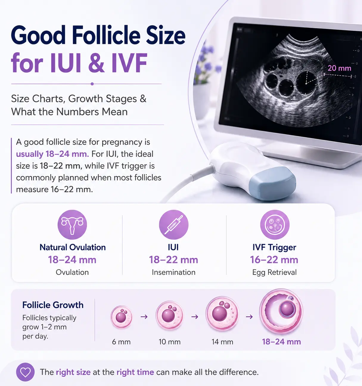
Good Follicle Size for IUI & IVF: Size Charts, Growth Stages & What the Numbers Mean
If you're trying to conceive naturally or undergoing IUI or IVF, you've likely wondered whether your follicle size is ideal. Follicle size isn't just a number on an ultrasound or on your report—it is one of the most trusted indicators fertility specialists use to asses

Fertility Supplements: A Complete Guide to Boosting Reproductive Health
Trying to conceive is often an emotional journey filled with hope, anticipation, and sometimes unexpected challenges. Many couples begin with lifestyle changes and healthy eating, while others require medical treatments such as ovulation induction, IUI, or IVF. Alongside

What Is Brain Fog? Symptoms and How to Get Rid of It
Brain fog describes a group of thinking and memory difficulties rather than a specific medical condition. You may find it harder to concentrate, remember information, organise tasks or find the right words. It can occur after poor sleep, stress, illness or changes in phy
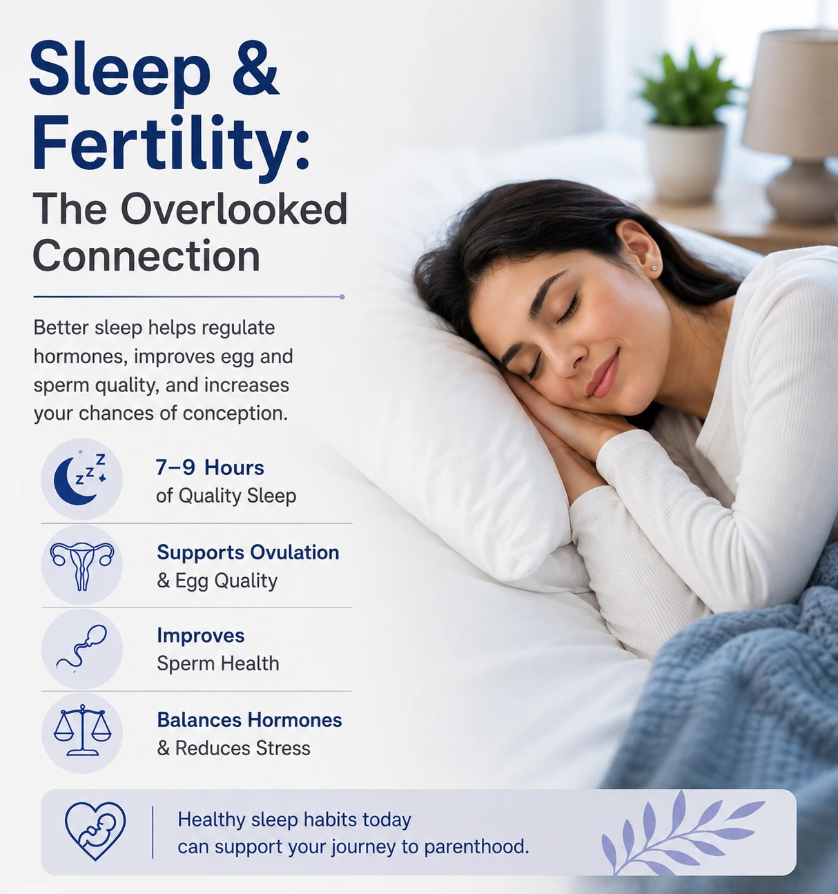
Can Poor Sleep Affect Fertility? Sleep, Hormones & Conception Explained
Sleep is a fundamental pillar of good health. It gives the body and mind time to repair, restore energy, regulate hormones, and maintain the normal functioning of cells, tissues, and organs, supporting overall physical and mental well-being. Sleep also plays a vital role i