
Thank You
Details submitted successfully
Our Customer Care Executive will reach out to you shortly.
Fetal Scan
A fetal scan monitors your baby's growth, heartbeat, position, and development during pregnancy. Learn when fetal scans are done, what they check, and what to expect.
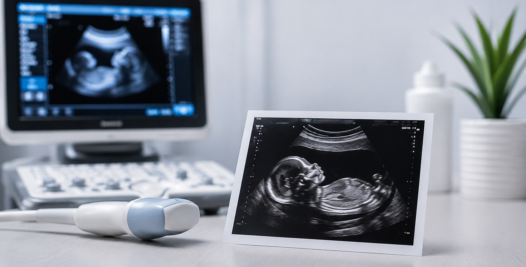
A fetal scan, also referred to as a fetal monitoring scan or fetal wellbeing scan, is a diagnostic ultrasound procedure performed during pregnancy to evaluate the health, growth, and development of the baby inside the womb. Using high-frequency sound waves, the scan creates real-time images of the fetus, placenta, and amniotic fluid — giving your doctor a clear picture of how your pregnancy is progressing at each stage.
Fetal scans are a routine and essential part of antenatal care. They are safe, non-invasive, and do not use radiation. Most expectant mothers undergo a series of fetal scans throughout pregnancy, each scheduled at a specific stage to assess different aspects of fetal development and maternal wellbeing.
The information gathered during a fetal scan helps your obstetrician track your baby's growth against expected milestones, identify any concerns early, and make informed decisions about your pregnancy care.
Here is an overview of when a fetal scan may be performed and what it is designed to assess:
1. First Trimester — Confirming the Pregnancy and Dating the Scan (6 to 14 weeks)
In the early weeks of pregnancy, a fetal scan is used to confirm that the pregnancy is developing within the uterus and that a heartbeat is present. This early scan helps establish the gestational age and estimated due date accurately, which is particularly important when periods are irregular or conception dates are uncertain.
During this stage, the scan also checks whether the pregnancy involves one baby or more, and assesses the development of the yolk sac and embryo at this early phase.
2. First Trimester Screening — Nuchal Translucency Assessment (11 to 14 weeks)
Between 11 and 14 weeks, a specialised fetal scan measures the fluid at the back of the baby's neck — known as the nuchal translucency. This measurement, combined with blood test results, helps assess the risk of chromosomal conditions such as Down syndrome (Trisomy 21), Edwards syndrome (Trisomy 18), and Patau syndrome (Trisomy 13). This scan forms part of the combined first trimester screening programme and is an important step in early risk assessment.
3. Second Trimester — Anomaly Scan (18 to 22 weeks)
Also referred to as the TIFFA scan (Targeted Imaging for Fetal Anomalies) or level 2 ultrasound, this is one of the most detailed fetal scans performed during pregnancy. It systematically evaluates the baby's anatomy from head to toe, examining the brain, spine, heart, kidneys, limbs, facial features, and abdominal organs. The scan also checks the position and appearance of the placenta and the amount of amniotic fluid surrounding the baby.
4. Third Trimester — Growth and Wellbeing Monitoring (28 weeks onwards)
As pregnancy progresses into the third trimester, fetal scans are performed to monitor the baby's growth and overall wellbeing. These scans assess the baby's estimated weight, head and abdominal measurements, limb length, amniotic fluid levels, placental position and grade, and fetal movements and breathing patterns observed during the scan. Serial growth scans may be recommended at regular intervals if there are concerns about slow growth, reduced fetal movements, or other risk factors.
5. Doppler Studies — Assessing Blood Flow
In certain pregnancies, particularly those considered high-risk, Doppler ultrasound is incorporated into the fetal scan. This technique measures blood flow through the umbilical cord, the baby's brain, and the placenta. Doppler studies help doctors assess whether the baby is receiving adequate oxygen and nutrients, and they play a key role in monitoring pregnancies complicated by conditions such as pre-eclampsia, gestational diabetes, or fetal growth restriction.
6. Late Pregnancy Scans — Position and Delivery Planning (36 weeks onwards)
Towards the end of pregnancy, a fetal scan is performed to confirm the baby's position — whether head-down (cephalic), breech, or transverse — which is important for planning the mode of delivery. This scan also re-evaluates the placental location, particularly to rule out low-lying placenta (placenta praevia), and checks amniotic fluid levels as part of fetal wellbeing assessment before delivery.
What Does a Fetal Scan Involve?
Most fetal scans are performed using an abdominal ultrasound, where a gel is applied to the abdomen and a handheld transducer is moved gently over the skin to capture images. In early pregnancy, a transvaginal scan — using a small probe placed gently inside the vagina — may provide clearer images of the developing embryo when the uterus is still low in the pelvis.
Fetal scans are generally comfortable and do not cause any harm to the mother or baby. The duration of the scan varies depending on the type and stage of pregnancy, typically ranging from 20 to 45 minutes. In some cases, if the baby is in an uncooperative position, you may be asked to walk around briefly or return for a repeat scan.
What If the Scan Shows Something Unusual?
Not every finding on a fetal scan signals a serious problem. Some observations may simply reflect a variation in fetal position, a temporary difference in measurements, or something that needs to be monitored over time. If a concern is identified, your doctor will explain what has been found, what it may mean, and what steps are recommended — which may include a repeat scan, a referral to a fetal medicine specialist, or additional investigations.
Early identification of any concern through regular fetal scanning allows for timely support, closer monitoring, and better pregnancy outcomes.
It is important to interpret fetal scan findings alongside your complete medical history, symptoms, and other test results. All decisions about further evaluation or care should be made in consultation with your obstetrician or fetal medicine specialist.